MAIN & TEXT MESSAGE

281.440.0101

MAIN & TEXT MESSAGE

281.440.0101

google translator

32 years

Best ultrasound scan


Top ultrasound imaging

ULTRASOUND

Ultrasound is a procedure that uses high-frequency sound waves to view internal organs and produce images of the human body. The human ear cannot hear the sound waves used in an ultrasound. Ultrasound is:


  • Noninvasive, which means it does not penetrate the skin or body openings, and
  • Diagnostic, which means it is used to determine what disease or condition is present


The technical term for ultrasound imaging is sonography.  Ultrasound technology was originally developed as a sonar to track submarines during World War I. It was first used medically in the 1950s and is considered very safe.  The original ultrasound scanners produced still images, but modern scanners produce moving pictures, which are easier to interpret.
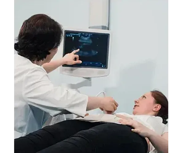
ULTRASOUND

How Does An Ultrasound Work?


Ultrasound imaging uses the principles of sonar developed for ships at sea. As sound passes through the body it produces echoes, which can identify distance, size and shape of objects inside.
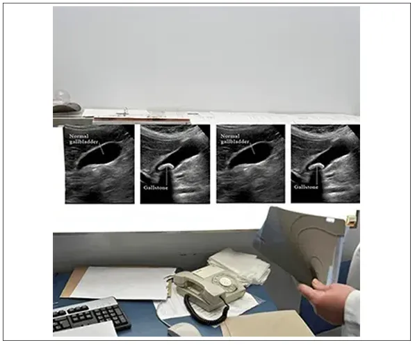
 

  • During the ultrasound examination, a machine called a transducer is used to view the target organ and produce pictures for study. The transducer emits sound and detects the returning echoes when it is placed on or over the body part being studied.
  • When the emitted sound encounters a border between two tissues that conduct sound differently, some of the sound waves bounce back to the transducer, creating an echo.
Ultra sound Treatment

  • The echoes are analyzed by a computer in the ultrasound machine and transformed into moving pictures of the organ or tissue being examined.


  • Ultrasound waves pass easily through fluids and soft tissues, making the procedure especially useful for examining fluid-filled organs such as the uterus in pregnancy, as well as the gallbladder, and soft organs like the liver.


  • Ultrasound waves are unable to penetrate bone or gas, so ultrasound is of limited use for examining regions surrounded by bone, or areas that contain gas or air. Even so, ultrasound has been used to examine most parts of the body.

Need To Know

Getting your results

Your ultrasound images will be analyzed by a radiologist, a physician who specializes in ultrasound and other radiology testing. The radiologist will send a signed report which includes an interpretation of the image to your primary physician. You may receive your results right after your scan. If not, you will receive information on the report through your primary care physician.

Nice To Know

What are the benefits and limitations of ultrasound?


  • Ultrasound is a noninvasive imaging technique. It is a painless procedure.
  • Ultrasound is widely available, low cost and easy to use.
  • Because it does not use radiation, the side effects of radiation are not an issue. So, ultrasound is the preferred technique for monitoring pregnant women and their unborn children.
  • Real-time images are generated by ultrasound, so it is a good tool for guiding invasive procedures like needle biopsies.
  • Ultrasound can display the movement and actual function of the body's organs and blood vessels.
  • There are no known harmful effects of standard ultrasound imaging.
  • The main limitation of ultrasound imaging is that it does not reflect clearly from bone or air. Therefore, other imaging techniques are preferred for areas such as the lungs and the bones.


How Do You Prepare For An Ultrasound Scan?


No special preparation is required for a routine ultrasound. Wear loose comfortable clothing to your ultrasound appointment. For a liver or gallbladder scan, the patient is usually asked to fast (take nothing by mouth) for several hours before the test.


What Happens During An Ultrasound Procedure?


You will probably be asked to lie down on a bed or table for the scan. Clothing over the area to be scanned is removed, and a special warm oil or gel is applied to the skin. This is to achieve good contact as the transducer is passed back and forth.


Ultrasonic waves are inaudible and cause no sensation, though pressure from the transducer may be uncomfortable. The scan usually takes about 15 minutes. During the procedure, you will probably be able to watch the ultrasound images on the screen attached to the scanner.


When a scan is performed in conjunction with a biopsy, a local anesthetic reduces or eliminates any discomfort.


Normal activities can be resumed immediately after the test. Ultrasound is very safe and painless, so there is little risk.